Figure 5.
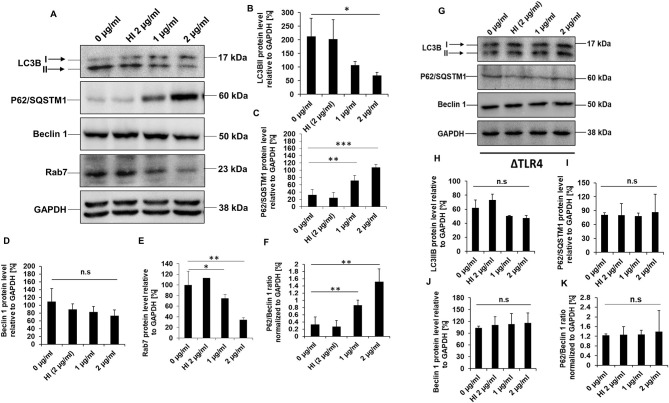
RipA inhibits autophagy in RAW264.7 cells in a TLR4 dependent manner. (A) Western blots demonstrating the inhibitory effect of RipA on cellular autophagy of RAW264.7 cells using autophagy markers. RAW264.7 cells were treated with RipA (1 and 2 μg/ml) for 24 h. Samples were prepared, run in SDS-PAGE, proteins were transferred onto polyvinylidene difluoride (PVDF) membrane and probed with indicated antibodies. The size of the protein bands is indicated. (B–F) Densitometric quantification of the protein bands (LC3BII, Beclin 1, Rab7, P62, and the ratio of P62/Beclin 1) are shown. Protein levels were represented as [%] to GAPDH. (G) Western blots showing the levels of autophagy markers in TLR4 knockout cells. Untreated and HI treated cells were used as negative controls. GAPDH was used as a loading control. (H–K) Densitometric quantitation of LC3BII, P62, Beclin1, and the ratio of P62/Beclin1 in treated ΔTLR4 cells. Protein levels of autophagy markers in ΔTLR4 cells were expressed as [%] to GAPDH. n.s indicates not significant. Data are representative of 3 independent experiments and expressed as means ± SD. *p < 0.05, **p < 0.01, and ***p < 0.001 vs. controls. n.s, not significant.

