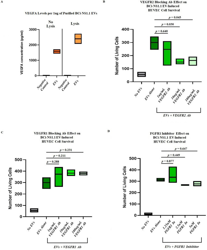
Figure 4.
VEGFR2, but not VEGFR1 or FGFR1, is involved in human airway basal cell EV–mediated survival of HUVECs. (A), Results of ELISAs, showing the presence of VEGFA in EVs isolated from BCi-NS1.1 cells. Negative control, an equal volume of a preparation from BEGM without BPE incubated for 48 h in flasks without cells and then processed by means of the same procedures used to isolate EVs. The floating bar graphs depict results from 3 batches of isolated EVs, with each batch run in duplicate. The horizontal lines inside the floating bars represent the mean concentrations from the 3 batches. The ELISAs were also performed after EVs were incubated with RIPA buffer to lyse them open. VEGFA was detected both with and without lysis. B–D, Floating bar graphs of the numbers of living HUVECs counted after 48-h incubation with either no EVs or 10 µg/mL EVs from BCi-NS1.1 cells in the presence of increasing concentrations of (B) VEGFR2-blocking antibody IMC-C11, (C) VEGFR1-blocking antibody IMC-18F, and (D) FGFR1 small-molecule inhibitor SSR128129E. The horizontal lines inside the floating bars represent the means of 3 experiments; adjusted P values correspond to differences between the bars on either end of the horizontal line below them and were calculated using Dunnett’s multiple comparisons test, with P ≤ 0.05 considered statistically significant.

