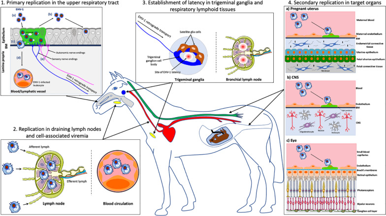
FIGURE 1.
Schematic representation of the pathogenesis of EHV-1 in the horse. (1) Primary EHV-1 replication in the epithelial cells of the upper respiratory tract: (a) EHV-1 infection (green); (b) Viral spread within the respiratory epithelium and viral shedding; (c) EHV-1 crosses the basement membrane (BM) and penetrates the lamina propria via the use of single infected leukocytes; (d) EHV-1 reaches the blood circulation and draining lymph nodes; (e) EHV-1 enters nerve endings of the peripheral nervous system and spreads in the retrograde direction to the trigeminal ganglia (TG). (2) EHV-1 replication in the draining lymph nodes and establishment of a cell-associated viremia in peripheral blood mononuclear cells (PBMC). (3) Establishment of EHV-1 latency in TG neurons and respiratory lymphoid tissues. (4) Via a cell-associated viremia in PBMC, EHV-1 is transported to target organs such as the pregnant uterus (4a), the central nervous system (CNS) (4b) or the eye (4c), where it initiates a secondary replication in the endothelial cells lining the blood vessels of this organ. gray, respiratory tract; red, blood circulation; yellow, lymph nodes; blue, TG; green, spinal cord. Drawings are partially based on SMART servier medical art templates.

