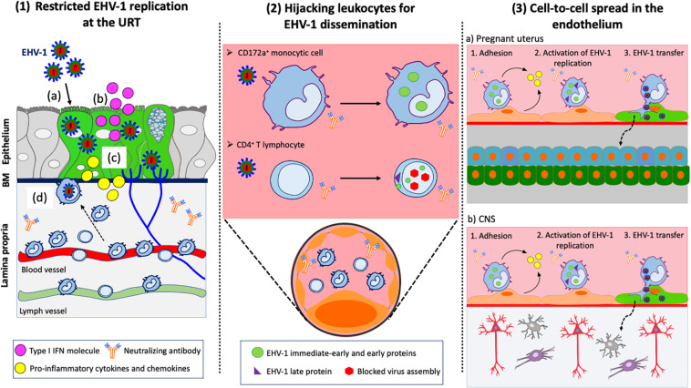
FIGURE 2.
EHV-1 immune evasive strategies. (1) Restricted replication at the URT: (a) EHV-1 infects respiratory epithelial cells; (b) Infected epithelial cells produce type I IFN to limit viral spread within the upper respiratory tract (URT); (c) Infected epithelial cells produce pro-inflammatory cytokines and chemokines to recruit immune cells to the site of infection; (d) CD172a+ cells and T lymphocytes migrate to the URT. EHV-1 uses these cells to cross the basement membrane (BM) and penetrate the lamina propria. (2) Hijacking leukocytes for viral dissemination. EHV-1 silences its replication in immune cells to avoid detection by neutralizing antibodies in the blood circulation and draining lymph nodes. EHV-1 only expresses immediate-early and early proteins in CD172a+ cells. All classes of EHV-1 proteins are expressed in T lymphocyte but viral assembly is blocked, and no progeny virions are released. (3) Cell-to-cell spread in the endothelium of target organs: EHV-1 infected immune cells are transported via the blood circulation to the pregnant uterus (a) and CNS (b). The adhesion of infected immune cells to the endothelial cells lining the blood vessels of target organs activates viral replication. New EHV-1 progeny virions are released from infected immune cells and transferred to endothelial cells via (micro)fusion events. Dashed arrows represent further spread of virus particles through connective tissues or CNS. Drawings are partially based on SMART servier medical art templates.

