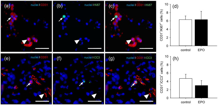
Figure 2.
Proliferating and apoptotic endothelial cells in MVF: (a–c) representative images of the immunohistochemical detection of CD31+/Ki67+ (arrows) and CD31+/Ki67− endothelial cells (arrowheads) of EPO-treated MVF within a CGAG matrix directly after seeding. Scale bars: 45 µm. (d) CD31+/Ki67+ endothelial cells (%) of vehicle- (control; white bar; n = 3) and EPO-treated (black bar; n = 3) MVF within CGAG matrices directly after seeding, as assessed by immunohistochemical analysis. Means ± SEM. (e–g) representative images of the immunohistochemical detection of CD31+/CC3+ (arrows) and CD31+/CC3− endothelial cells (arrowheads) of EPO-treated MVF within a CGAG matrix directly after seeding. Scale bars: 45 µm, and (h) CD31+/CC3+ endothelial cells (%) of vehicle- (control; white bar; n = 3) and EPO-treated (black bar; n = 3) MVF within CGAG matrices directly after seeding, as assessed by immunohistochemical analysis.
Means ± SEM.

