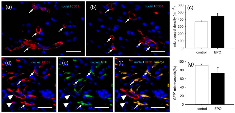
Figure 6.
Vascularization of implanted MVF-seeded CGAG matrices: (a, b) immunohistochemical detection of CD31+ microvessels (arrows) in MVF-seeded CGAG matrices on day 14 after implantation into full-thickness skin defects within the dorsal skinfold chamber of a vehicle- (a) and an EPO-treated (b) C57BL/6 mouse. Scale bars: 30 µm, (c) microvessel density (mm−2) of MVF-seeded matrices in vehicle- (control; white bar; n = 8) and EPO-treated (black bar; n = 8) C57BL/6 mice on day 14 after implantation, as assessed by immunohistochemical analysis. Means ± SEM, (d–f) immunohistochemical detection of CD31+/GFP+ microvessels (arrows) and CD31+/GFP− microvessels (arrowheads) in a MVF-seeded CGAG matrix on day 14 after implantation into a full-thickness skin defect within the dorsal skinfold chamber of an EPO-treated C57BL/6 mouse. Scale bars: 30 µm, and (g) GFP+ microvessels (%) in MVF-seeded matrices of vehicle- (control; white bar; n = 8) and EPO-treated (black bar; n = 8) C57BL/6 mice on day 14 after implantation, as assessed by immunohistochemical analysis.
Means ± SEM.

