Figure 2.
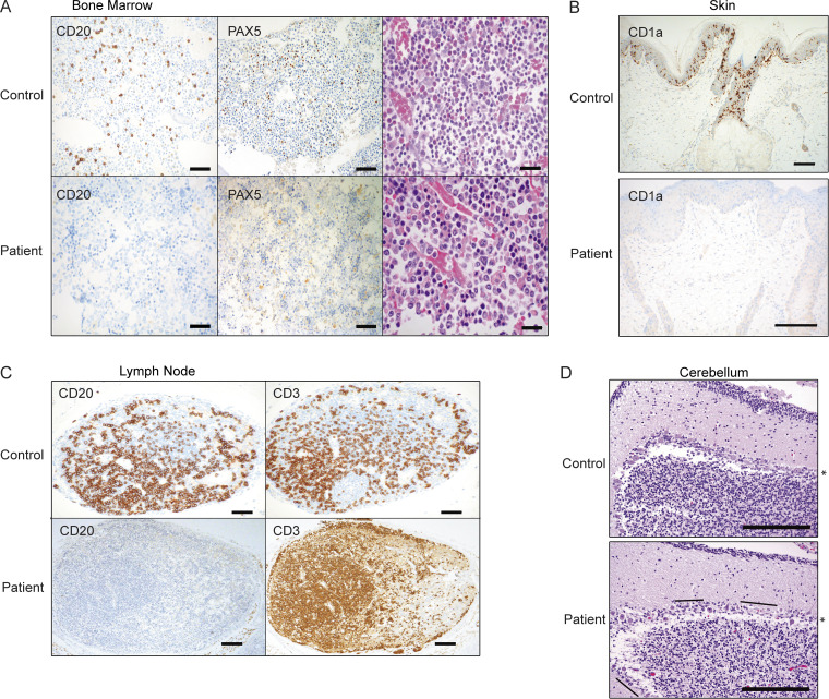
Histological evidence of selective cytopenias. Biopsy or autopsy samples were analyzed and compared with cases controlled for equal stress or treatments. Data are representative. (A) Proband BM with left-shifted myelopoiesis and similar cellularity as control BM. All cell lines are present, although megakaryocytes are decreased. Immunohistochemical stains (CD20 and PAX5) confirm the presence of scattered B cells in the control sample but none in the patient (diaminobenzidine, 100×; scale bar, 200 µm; H&E, 200×; scale bar, 50 µm). (B) Langerhans cells are highlighted with CD1a in the epidermis of the control skin sample and absent in the patient (diaminobenzidine, 100× [top] and 200× [bottom]; scale bars, 100 µm). (C) Control lymph node has abundant CD20+ and CD3+ lymphocytes, with few primary follicles. The CD3+ T cell population is intact in the patient, but no B cells are identified with CD20 (diaminobenzidine, 40×; scale bar, 300 µm). (D) Cerebellum sections were stained with H&E, highlighting the layer of confluent Purkinje cells (*) in a control subject and regions of Purkinje cell loss (lines) in the patient (200×; scale bars, 100 µm).

