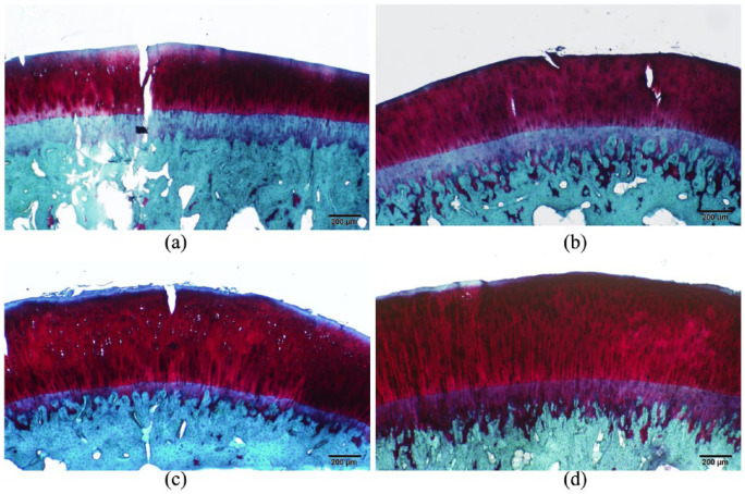
Figure 6.
Histopathological images of the tibial femoral condyle cartilage: The control (A) demonstrated fissuring and reduction of safranin-O staining. The sham (B) and high-dose (D) groups were nearly indistinguishable, representing normal articular cartilage. The low-dose group (C) exhibited mild fissuring.

