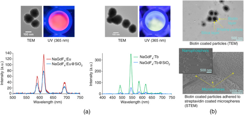
Fig. 2.
(a) Luminescence spectra of (red) and (green) nanophosphors, TEM images above the spectra show silica coated particles. (b) TEM image of biotin-coated nanophosphors (top), and STEM image of biotin-coated nanophosphors adhered to streptavidin coated microspheres (bottom), inset image is a zoomed-in view.

