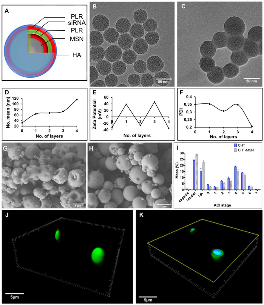
Figure 1.
Synthesis and characterization of aerosolized LbL nanoparticles. (A) Schematic representation siRNA-containing LbL nanoparticles. TEM images of (B) MSN’s loaded with IR-780 dye and (C) siRNA LbL nanoconstructs from MSN cores, scale bars 50nm. (D) hydrodynamic diameter, (E) zeta potential, and (F) polydispersity index (PDI) of the LbL nanosystems. SEM images of the chitosan microparticles containing LbL nanoparticles in A-F: (G) CHT (x 16,000); (H) CHT-LbL siRNA (x 30,000), scale ba–s - 1m. (I) Powder dispersion among the micronized powders by ACI. 3D reconstruction of CHT-LbL siRNA micronized powders and (J) their cross-section image (K). Green: AF 488 siRNA (λexc = 473 nm; λemm = 520 nm); Blue – DAPI, CHT autofluorescence (λexc = 405 nm; λemm = 461 nm), scale bar - 5μm.

