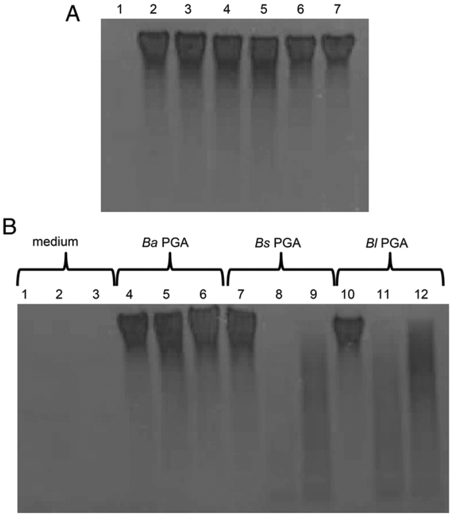
FIGURE 8.

B. anthracis PGA persists longer in high m.w. form in monocyte and iDC culture media than B. subtilis PGA or B. licheniformis PGA. (A) 125 μg/ml B. anthracis PGA, B. licheniformis PGA, or B. subtilis PGA was incubated in culture medium without cells for 7 d at 37°C/5%CO2. Gel order is as follows: 1) culture medium, 2) day 0 B. anthracis PGA, 3) day 7 B. anthracis PGA, 4) day 0 B. subtilis PGA, 5) day 7 B. subtilis PGA, 6) day 0 B. licheniformis PGA, and 7) day 7 B. licheniformis PGA. (B) 125 μg/ml B. anthracis PGA, B. licheniformis PGA, or B. subtilis PGA was added to cultures of human monocytes or iDCs. Samples of the culture media were collected after three (monocytes) or two (iDCs) days of incubation. Gel order is as follows: 1) culture medium, 2) spent monocyte culture medium, 3) spent iDC culture medium, 4) B. anthracis PGA alone, 5) B. anthracis PGA with monocytes, 6) B. anthracis PGA with iDCs, 7) B. subtilis PGA alone, 8) B. subtilis PGA with monocytes, 9) B. subtilis PGA with iDCs, 10) B. licheniformis PGA alone, 11) B. licheniformis PGA with monocytes, and 12) B. licheniformis PGA with iDCs. Media samples were electrophoresed on gradient gels and stained with Stains-All. Four experiments were conducted with cells from four donors. Data from a single representative experiment are shown. Ba, B. anthracis; Bl, B. licheniformis; Bs, B. subtilis.
