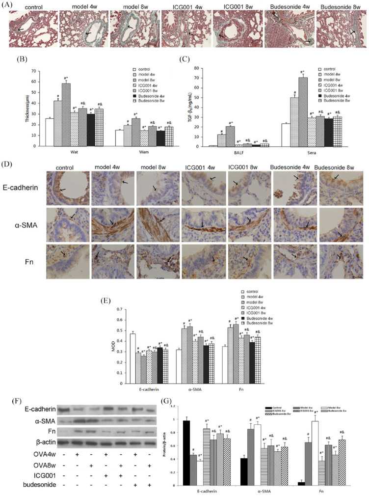
Figure 2.
ICG001 attenuated airway remodeling and inhibited transforming growth factor (TGF)-β1-induced epithelial mesenchymal transition(EMT) in a rat model of asthma. (A) Collagen deposition in the subepithelial fibrotic tissues of all seven groups, analyzed using Masson’s trichrome stain (magnification, ×100). Black arrows indicate collagen deposition in airways. (B) The thickness of the airway wall (Wat) and the airway smooth muscle (Wam) in all seven groups. (C) The levels of TGF-β1 in the bronchoalveolar lavage fluid (BALF) and sera of all seven groups. (D) Immunohistochemistry images of E-cadherin, alpha-smooth muscle actin (α-SMA), and fibronectin staining in pulmonary tissue slices of all seven groups (magnification, ×400). Black arrows indicate E-cadherin, α-SMA, and fibronectin (Fn) expressions in airways. (E) Quantitative analysis of the immunohistochemistry results of E-cadherin, α-SMA, and Fn, based on relative densitometry intensity. (F) Western blot analysis for E-cadherin, α-SMA, and Fn in the pulmonary tissue of all seven groups. (G) Quantitative analysis of the Western blot results of E-cadherin, α-SMA, and Fn, based on relative densitometry intensity.
MOD: mean optical density.
The values are expressed as mean ± standard deviation (n = 8).
#p < 0.01 versus control group.
*p < 0.01 versus 4 weeks (4w) model group.
&p < 0.01 versus 8 weeks (8w) model group.

