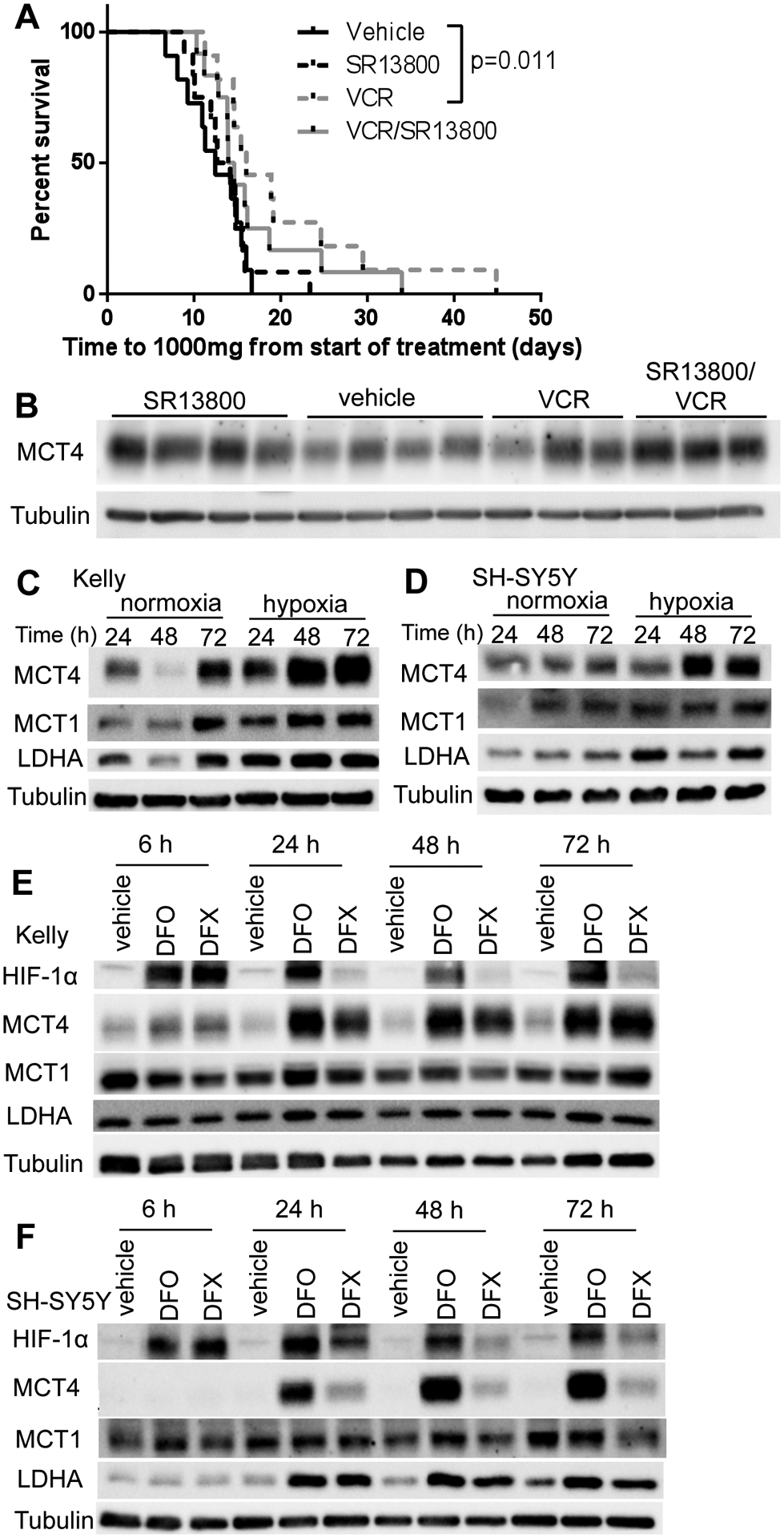
Figure 6. MCT4 expression is elevated in neuroblastoma under conditions of hypoxia and HIF-1α induction.

(A) Response of Kelly xenografts in BALB/c nude mice to vehicle, SR13800 (30 mg/kg), vincristine (VCR, 0.2 mg/kg) and VCR/SR13800. (B) Tumor protein levels of MCT4 harvested at end point. (C–D) MYCN-amplified Kelly (C) and MYCN non-amplified SH- SY5Y (D) cells were incubated in normoxic or hypoxic conditions for 24, 48 and 72h and samples were analysed by Western blot for MCT1, MCT4 and LDHA. Kelly (E) and SH-SY5Y (F) were treated with HIF inducers desferrioxamine (DFO, 20mM) or deferasirox (DFX, 100mM) over a timecourse (6h–72h) and levels of HIF1α, MCT1, MCT4 and LDHA were analysed by Western blot. Tubulin served as a loading control. Representative blots from three independent experiments.
