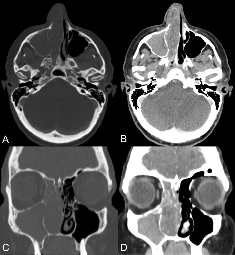
Fig. 1.
CT scan of nose and paranasal sinuses. a Bone demineralization of ethmoidal cells and the medial maxillary sinus wall in axial section with a bone window. b Soft-tissue-density mass occupying the entire right nasal cavity in axial section with a soft-tissue window. c Bone demineralization of ethmoidal cells, turbinates, and the uncinate process in coronal section with a bone window. d Opacification of the maxillary sinus in coronal section with a soft-tissue window

