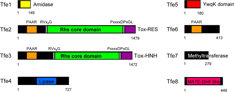
Figure 3.
Domain composition of T6SS-related effectors present in P. fluorescens F113. The domain organization of the putative effectors is shown, with amidase domain in yellow, PAAR motifs indicated in orange, RVxxxxxxxxG and PxxxxDPxGL motifs in grey, Rhs domains in green, HNH nuclease motifs (Tox-HNH and Tox-SHH) in purple, YwqK domain in red and MATE domain in pink. Structural-based homology prediction was determined using the Protein Homology/analogy Recognition Engine (Phyre) server.

