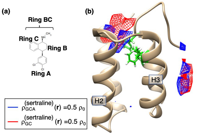
Figure 4.
(a) Chemical structure of sertraline, where ring A and ring BC (rings A and B) are shown. (b) Spatial density (blue-colored contours) for the geometric center of Ring A of sertraline in the sertraline–mSin3B system, where the contour level is (). Red-colored contours are , which is spatial density of the geometric center of the entire sertraline. Labels H2 and H3 represent helices 2 and 3, respectively. Green-colored residues are Val 75, Phe 93, and Phe 96 of mSin3B. See Supplementary Section 4 for procedure to calculate .

