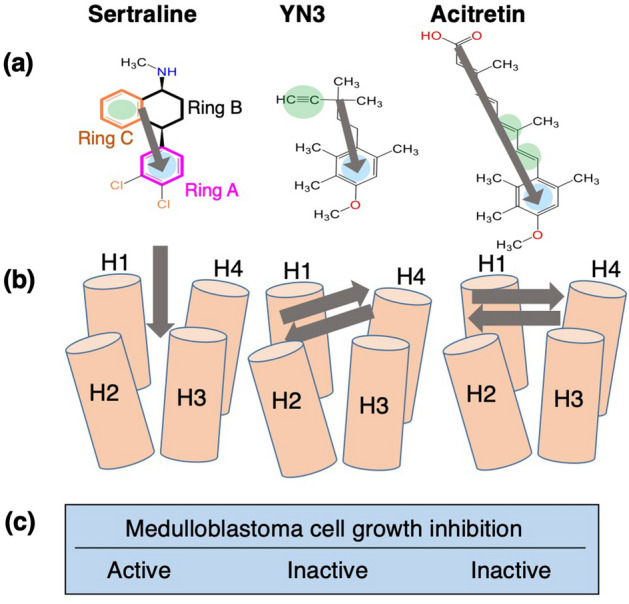
Figure 8.

(a) Chemical structures of three compounds, and their orientations (gray arrows). Rings A, B and C of sertraline are depicted in magenta, black, and orange, respectively. Blue circles represent aromatic rings that are expected to interact with the hydrophobic cleft of mSin3B from conventional structure–activity relationship (SAR). Green circles represent -electron rich regions. (b) Orientations of the compounds in the bound forms with mSin3B, resulted from the present MD simulation study. (c) Presence/absence of medulloblastoma cell-growth inhibition activities for the compounds.
