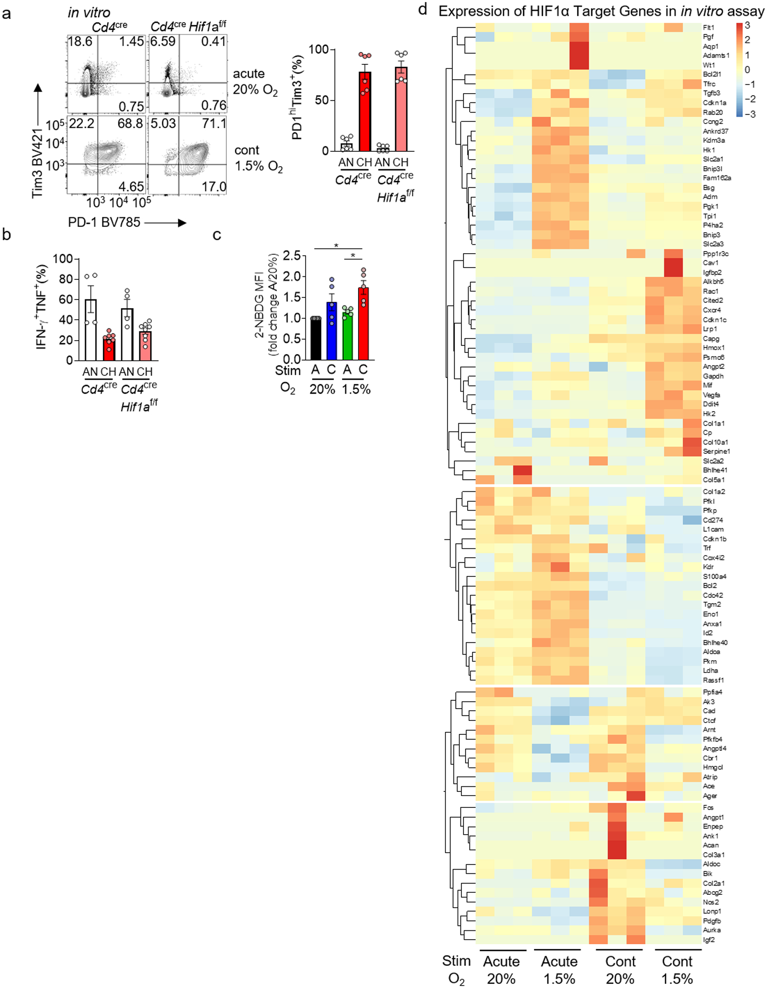
Extended Figure 4. HIF-1α is dispensable for continuous stimulation under hypoxia-induced dysfunction, although hypoxia signaling is active in response to hypoxia.

a, PD-1 and Tim-3 staining in WT or HIF-1α-deficient CD8+ T cells continuously stimulated under hypoxia as in Fig. 2. each group n=6. b, Cytokine production of restimulated WT and HIF-deficient T cells as in a. WT AN n=4, CH n=7; HIF-deficient AN n=4, CH n=8. c, Quantification of 2-NBDG staining as in Figure 2. each group n=5. d, Heatmap of known HIF-1α target genes from the transcriptional analyses in Fig. 3. Each group n=3. All data are representative of 3–6 independent experiments. *p < 0.05 by one-way ANOVA with Dunnett’s multiple comparison test (c). Error bars indicate SEM.
