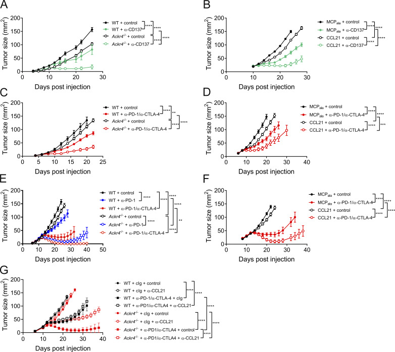
Figure 5.
ACKR4 deficiency enhances responsiveness to immunotherapy. (A) Tumor growth in WT or Ackr4−/− mice injected with 105 E0771 cells and administered 100 µg anti-CD137 (clone 3H3) or rat IgG every 3 d from days 10 to 19; n = 7–9. (B) Tumor growth in WT mice injected with 105 E0771 cells and administered MCPala or CCL21 (3 µg intratumorally) plus 100 µg anti-CD137 or rat IgG every 3 d from days 10 to 19; n = 5. (C) Tumor growth in WT or Ackr4−/− mice injected with 105 B16F10 cells and administered 250 µg anti–PD-1 (clone RMP1-14) and 250 µg anti–CTLA-4 (clone UC10-4F10) or 500 µg control rat/hamster Ig every 3 d from days 6 to 15; n = 5–6 mice. (D) Tumor growth in WT mice injected with 105 B16F10 cells and administered MCPala or CCL21 (3 µg intratumorally) plus 250 µg anti–PD-1 and 250 µg anti–CTLA-4 or 500 µg control rat/hamster Ig every 3 d from days 6 to 15; n = 6. (E) Tumor growth curves from WT or Ackr4−/− mice injected subcutaneously with 105 MC38 cells and administered 250 µg anti–PD-1, 250 µg anti–PD-1/250 µg anti–CTLA-4, or 500 µg rat/hamster Ig every 3 d from days 12 to 21; n = 7–8 mice. (F) Tumor growth curves from WT mice injected subcutaneously with 105 MC38 cells and administered MCPala or CCL21 (3 µg, intratumorally) plus 250 µg anti–PD-1 and 250 µg anti–CTLA-4 or 500 µg rat/hamster Ig every 3 d from days 12 to 21; n = 10 mice. (G) Tumor growth curves from WT or Ackr4−/− mice injected subcutaneously with 105 MC38 cells and administered 250 µg anti-PD-1 (clone RMP1-14) and 250 µg anti–CTLA-4 (clone UC10-4F10) or 500 µg rat/hamster Ig every 3 d from days 12 to 21 plus control or anti-CCL21 (100 µg) every 6 d from day −1; n = 6–7 mice. Data are representative from two independent experiments (A–F) or are from one experiment (G). All statistical analyses were performed using a two-way ANOVA, and data are presented as mean ± SEM. **, P ≤ 0.01; ***, P ≤ 0.001; ****, P ≤ 0.0001.

