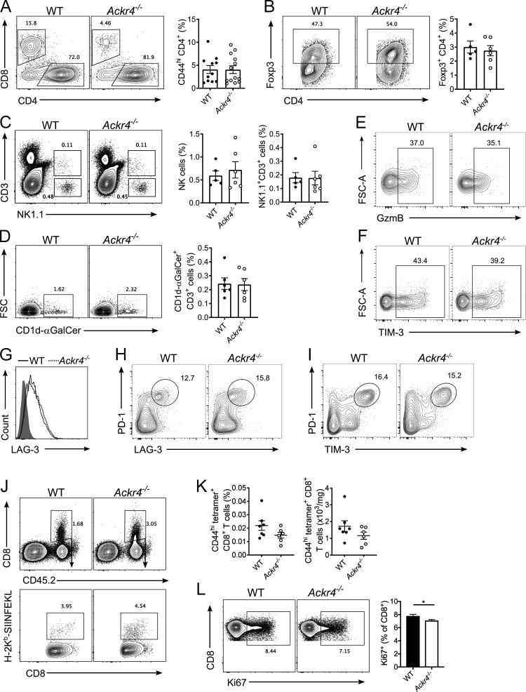
Figure S1.
Tumor-infiltrating lymphocytes and CD8+ T cell priming in mice bearing E0771 tumors. (A–G) WT or Ackr4−/− mice were injected with 105 E0771 cells into the fourth mammary gland and analyzed 18–21 d after injection. (A) Representative gating and frequency (of total viable cells) of intratumor CD44hi CD4+ T cells; n = 11–13. (B) Representative gating and frequency of intratumor Foxp3+ regulatory t cells. n = 5–6. (C) Representative gating and frequency of intratumor NK cells and NK1.1+ CD3+ NKT cells. n = 5–6. (D) Representative gating and frequency of type I invariant NKT cells. n = 5–6. FSC, forward scatter. (E–H) Representative gating strategy of intratumor CD8+ T cells for (E) granzyme B, (F) TIM-3, (G) LAG-3, (H) PD-1+ LAG-3+, and (I) PD-1+ TIM-3+ (data in Fig. 2, C and E–G). (J) Representative gating strategy of OVA-specific CD8+ T cells in E0771-OVA tumors (data in Fig. 2 F). (K) Frequency and number of OVA-specific CD44hi CD8+ T cells in dLNs of mice injected with 5 × 10⁵ E0771-OVA cells; n = 6–7. (L) Representative gating and frequency of Ki67 expression on CD8+ T cells in the dLNs of mice injected with 105 E0771 cells. n = 10, unpaired t test. Data representative of at least two independent experiments. Data are presented as mean ± SEM. *, P ≤ 0.05.

