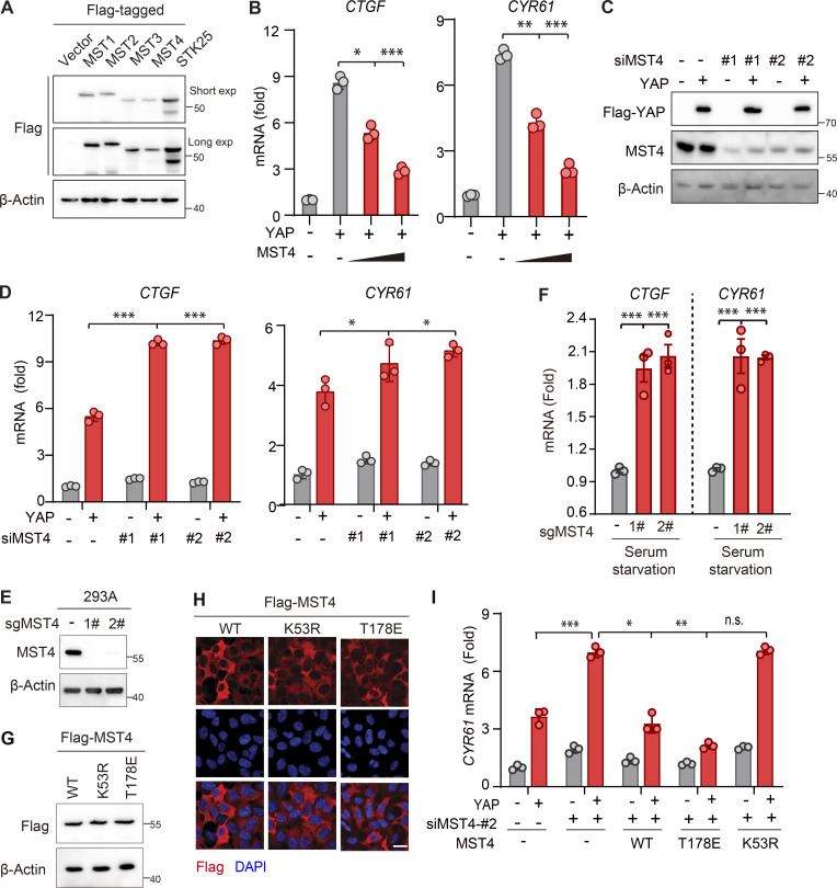
Figure S1.
MST4 negatively regulates YAP activation. (A) Western blotting analysis of exogenously expressed indicated constructs using anti-Flag antibodies (n = 2). (B) MST4 dose-dependently inhibits YAP target gene expression. HEK293FT cells were treated as described in Fig. 1 B followed by qPCR analysis of CTGF and CYR61 mRNA expression (n = 3, *, P < 0.05; **, P < 0.01; ***, P < 0.001, one-way ANOVA with Dunnett’s post hoc analysis, compared with column 2). (C) Validation of the siRNA-mediated MST4 knockdown efficiency by Western blotting using indicated antibodies (n = 2 biological repeats). (D) MST4 depletion activates YAP target gene expression. HEK293FT cells were treated as described in Fig. 1 C followed by qPCR analysis of CTGF and CYR61 mRNA expression (n = 3, *, P < 0.05; ***, P < 0.001, one-way ANOVA with Dunnett’s post hoc analysis, compared with column 2). (E) Generation and validation of the 293A MST4 KO cells using CRISPR-Cas9 technology. (F) MST4 depletion promotes YAP target gene expression. 293A cells (WT and KO) were treated with serum-free medium for 12 h. mRNA was extracted, and transcriptional levels of CTGF and CYR61 were examined by real-time qPCR (n = 3, ***, P < 0.001, one-way ANOVA with Dunnett’s post hoc analysis, compared with control gRNA). (G and H) Generation and validation of the 293A KO cells with reconstitution with indicated constructs as used in Fig. 1 G by either IFA (G; n = 1 biological repeat) or Western blotting (H; n = 1 biological repeat). (I) 293A cells pretreated with MST4 siRNA#2 were transfected with indicated MST4 constructs, together with or without YAP plasmid. qPCR analysis of CYR61 mRNA expression were performed in each treatment (n = 3, n.s., not significant, *, P < 0.05; **, P < 0.01; ***, P < 0.001, one-way ANOVA with Dunnett’s post hoc analysis, compared with column 4). Data are presented as the mean ± SEM. Related to Fig. 1.

