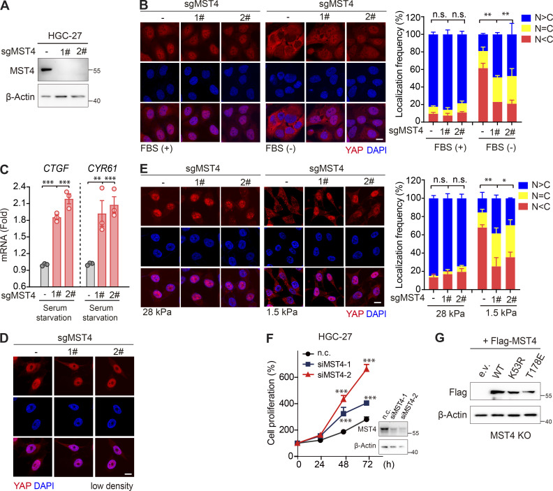
Figure S4.
MST4 limits YAP activation and cell proliferation in GC cells. (A) Generation and validation of HGC-27 MST4 KO cells using CRISPR-Cas9 technology. (B) MST4 KO promotes YAP nuclear localization in response to serum starvation. HGC-27 cells (WT and MST4 KO) were subjected to serum starvation for 12 h and processed to IFA assay using anti-YAP antibody (scale bar, 5 µm; n = 3, n.s., not significant, **, P < 0.01, one-way ANOVA with Dunnett’s post hoc analysis). (C) MST4 KO promotes YAP target gene expression. HGC-27 cells (WT and KOs) were treated with serum-free medium for 12 h. mRNA was extracted, and transcriptional levels of CTGF and CYR61 were examined by real-time qPCR (n = 3, **, P < 0.01; ***, P < 0.001, one-way ANOVA with Dunnett’s post hoc analysis, compared with control gRNA). (D) HGC-27 cells (WT and MST4 KO) at low cell density (10%) were subjected to IFA assay using anti-YAP antibody (scale bar, 5 µm). (E) HGC-27 cells (WT and MST4 KO) were seeded at either soft (1.5 kPa) or high stiffness (28.0 kPa) plates for 24 h before processing for IFA assay using anti-YAP antibodies (scale bar, 5 µm; n = 3, n.s., not significant, *, P< 0.05; **, P < 0.01, one-way ANOVA with Dunnett’s post hoc analysis, compared with control gRNA). (F) Knockdown of MST4 stimulates GC cell growth. HGC-27 cells pretreated with indicated siRNAs were seeded at 1,000 cells/well density, and cell proliferation curves over the indicated time were measured by CellTiter Luminescent-based assay (four replicates per cell line for three repeats, ***, P< 0.001; two-way ANOVA with Tukey’s post hoc test). (G) Western blotting analysis of the reconstitution of MST4 constructs in HGC-27 MST4 KO cells used in Fig. 5 H. Data are presented as the mean ± SEM. n.c., non-targeting control. Related to Fig. 5.

