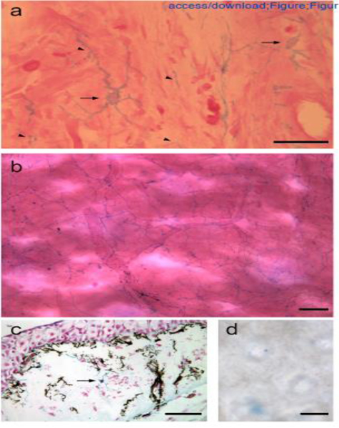
Figure 1.

Alcian blue-positive GRID cells in the loose connective tissue of the dermis.
High magnification (a) and low magnification (b) images of whole-mount preparations of dermal tissue illustrating the dendritic morphology of GRID cells that form an interspersed cellular network. (c) GRID cells (arrow indicates an alcian blue-positive cell body) are distinct from melanocytes (black cell bodies and processes). (d) Nitrous acid treatment degraded HS, and alcian blue-positive GRID cells were no longer evident in dermal tissues. Examples of GRID cell bodies are indicated by arrows, and expanded regions of GRID cell processes are indicated by arrowheads (a). Scale bars: 50 μm (a, d), 100 μm (b, c).
