Figure 5.

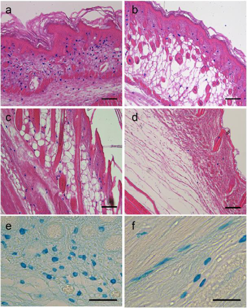
Alcian blue-positive cells in the connective tissues of mouse skin.
Alcian blue-positive cells were abundant throughout the dermal connective tissue of mouse skin at postnatal day 1 (a, PN1). These cells were observed at progressively decreasing abundance as the neonatal skin matured (b, PN6; c, PN9; and d, adult). The histology sections are oriented with the epidermis of the skin toward the upper-right of the images (a – d). At higher magnification without eosin counterstaining, the cell bodies were mostly round (e, PN1), although some appeared more elongate and stellate (f, PN9). Scale bars: 100 μm (A, B, C, D), 50 μm (E, F).
