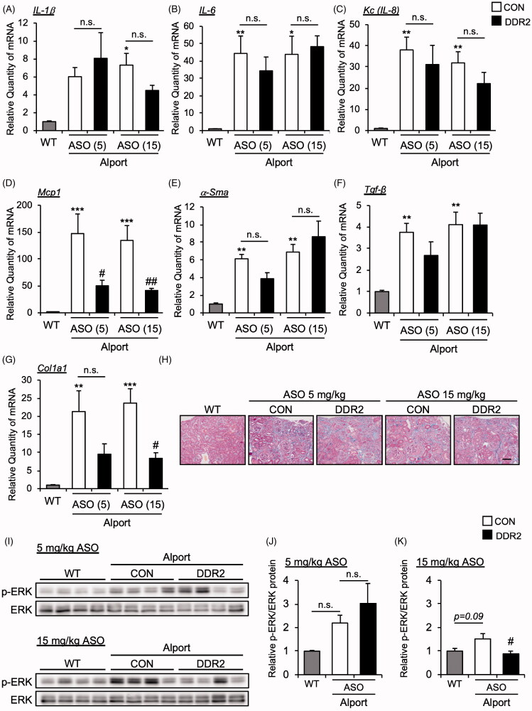
Figure 4.
DDR2 ASO did not affect inflammatory and fibrosis gene expression in Alport mice. Total RNA was isolated from kidney tissues of 24-week-old mice. Quantitative RT-PCR was performed to evaluate the expression of the indicated (A-D) cytokines and (E-G) renal fibrosis markers. The data were normalized to Gapdh. Bars indicate the mean ± S.E. (n = 5–6). *p<.05, **p<.01, ***p<.001 vs WT; #p<.05, ##p<.01 vs CON ASO, assessed by Dunnett’s test. (H) Masson-Trichrome staining of renal section of 24-week-old mice was performed. Scale bar, 100 µM. (I) Immunoblots of protein lysates from whole kidneys of WT and AS mice were probed with phosphorylated ERK (p-ERK) or ERK antibodies. (J,K) The p-ERK expression was quantified by multi gauge software and normalized with basal ERK. Bars indicate the mean ± S.E. (n = 4). #p<.05 vs CON ASO, assessed by Dunnett’s test.

