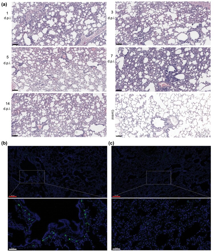
Figure 4.
Histopathological lung changes in tree shrews infected with HAdV-55 at 1, 3, 5, 7, and 14 d post-infection. (a) Haematoxylin and eosin (HE) staining of the lung tissues. (b) Immunofluorescence analysis of HAdV-55 in the lung tissues of HAdV-55-infected tree shrew at 3 d post-infection. (c) Immunofluorescence analysis of HAdV-55 in lung tissue of control tree shrew treated with PBS. A mouse polyclonal antibody raised against HAdV-55 virions was used for HAdV-55-specific staining. Black scale bar, 100 μm; red scale bar, 200 μm; white scale bar, 50 μm.

