Figure 1.

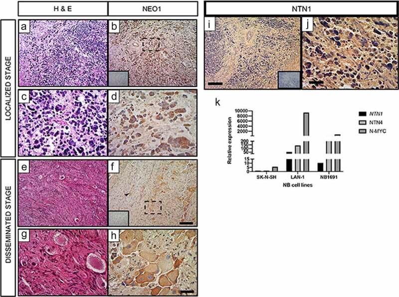
NEO1 is expressed in NB samples independently of tumoral stage and NTN1 has impaired expression. Immunohistochemically (IHC) analysis of NEO1 expression in NB samples. In all IHC Hematoxylin was used as a counterstaining a- d: Representative images of a NB patient sample classified at Localized Stage according to INRGSS. a, c: Hematoxylin-Eosin (H&E) staining, b: NEO1 expression (brown). Dotted square shows the area represented at higher magnification in d. e- h: Representative images of a NB patient sample classified at Disseminated Stage according to INRGSS. e, g: H&E staining, f, h: NEO1 expression. Dotted square shows the area represented in high magnification in h. Negative control of the antibody are shown as inset in b and f. Arrowhead indicates NEO1 staining in blood vessels. a, b, e, f: Low magnification Bar: 100 μm, c, d, g, h: High magnification Bar: 20 μm. Immunohistochemistry of NTN1 expression within NB. Representative light microscopy images of neuroblastoma samples from primary tumors. Hematoxylin was used for counterstaining. i, j: Representative images of a NB patient sample classified at Localized Stage according to INRGSS. i: Low magnification, j: High magnification. Negative control of each antibody is shown as an inset. Low magnification Bar: 100 μm, High magnification Bar: 20 μm. k: Relative expression of NTN1, NTN4 and N-MYC in indicated NB cell lines. GAPDH expression was used as housekeeping control. N = 21
