
Figure 5.
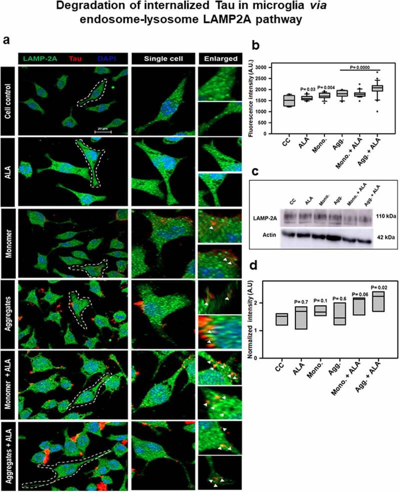
Degradation of internalized Tau in microglia via endosome–lysosome LAMP-2A pathway. Last step of degaradation pathway includes fusion of late endosome with lysosome studied by fluorescence microscopy. (a) Internalization of extracellular Tau (red) studied with its colocalization with LAMP-2A (green) after 24 h of ALA and extracellular Tau exposure. The images indicate levels of LAMP-2A and its colocalization with Tau. Enlarged panel indicates that specific area from the representative image showing colocalization of Tau and LAMP-2A, indicated with white triangles. Scale bar is 20 μm. (b) Intracellular intensity of LAMP-2A was calculated from immunofluorescence images and plotted as mean intensity inside the cell. The significance is P < 0.05. (c) Levels of LAMP-2A were detected by western blot after ALA and Tau exposure. (d) Quantification of intensity of protein bands, normalized with the β-actin as a loading control