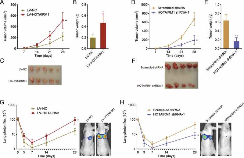
Figure 4.

The roles of HOTAIRM1 in ATC growth and metastasis in vivo. (A-C) HOTAIRM1 overexpressed or control 8505 C cells were subcutaneously inoculated into nude mice. Subcutaneous tumours volumes were measured every 7 days (A). At the 28th day after inoculation, subcutaneous tumours were resected, weighed (B), and photographed (C). (D-F) HOTAIRM1 depleted or control FRO cells were subcutaneously inoculated into nude mice. Subcutaneous tumours volumes were measured every 7 days (D). At the 28th day after inoculation, subcutaneous tumours were resected, weighed (E), and photographed (F). (G) Luciferase-labelled HOTAIRM1 overexpressed or control 8505 C cells were injected into tail vein of nude mice, followed by bioluminescence imaging to track lung metastasis. Represent images of mice at the 28th day after injection were shown. (H) Luciferase-labelled HOTAIRM1 depleted or control FRO cells were injected into tail vein of nude mice, followed by bioluminescence imaging to track lung metastasis. Represent images of mice at the 28th day after injection were shown. Data were shown as mean ± standard deviation of five mice in each group. *P < 0.05, **P < 0.01 by Mann-Whitney test
