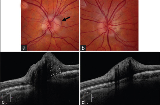
Figure 1.

Fundus photograph of the right eye (a) and the left eye (b)centered on the optic nerves show tilted optic discs with mild superonasal elevation in both eyes and mild swelling of the right optic nerve head. In the right eye, a peripapillary hemorrhage (black arrow) is seen along the superonasal margin of the disc. (c) Enhanced depth imaging-optical coherence tomography centered on the optic nerve shows a hyperreflective area (white outline) adjacent to the optic nerve head, suggesting a choroidal neovascular membrane. (d) Enhanced depth imaging-optical coherence tomography centered on the left optic nerve was normal.
