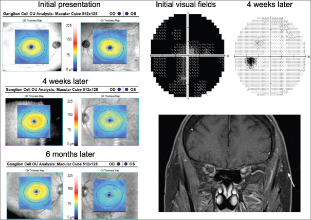
Figure 2.
Left idiopathic optic neuritis. Initial visual fields were diffusely depressed with full recovery after 4 weeks. However, on optical coherence tomography, early ganglion cell–inner plexiform layer thinning is seen. Despite visual field improvement, there was significant ganglion cell–inner plexiform layer thinning at 6 months

