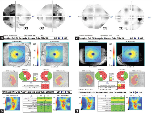
Figure 6.
Bitemporal hemianopia secondary to prolactinoma. (a) Optical coherence tomography of the ganglion cell–inner plexiform layer showing binasal thinning while the (b) retinal nerve fiber layer showing temporal thinning. (c) After medical treatment, the visual field defect resolved, but (d) the binasal ganglion cell–inner plexiform layer thinning persisted

