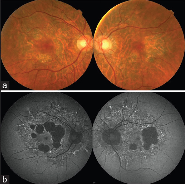
Figure 2.

(a) A 55-year-old female with the m.3243A>G mutation and maternally inherited diabetes and deafness. Color fundus photographs show discontinuous circumferential perifoveal atrophy that is typical of macular pattern dystrophy. (b) Large patches of hypo-autofluorescence correspond to the perifoveal areas of macular atrophy. There is diffuse, speckled autofluorescence that extends beyond the temporal arcades that was not apparent on color fundus photographs.
