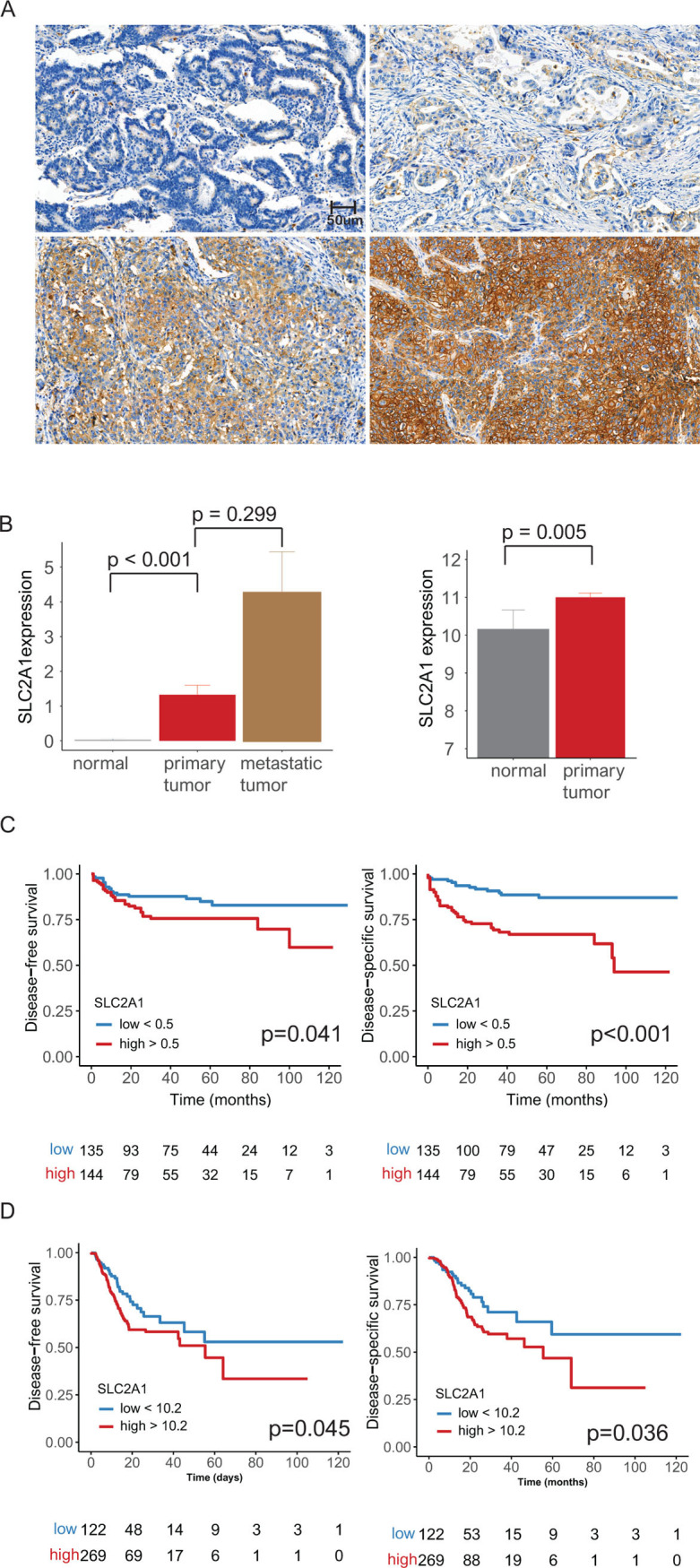
Fig 1.

(A) Representative microphotographs showing negative (top left), weak (top right), moderate (bottom left) and strong intensity (bottom right) SLC2A1 expression in gastric adenocarcinoma by immunohistochemical staining (original magnification ⅹ400). (B) Bar plots of SLC2A1, Eulji Hospital cohort paired (matched) samples: SLC2A1 expression was highest in metastatic tumors followed by primary tumors (left). TCGA paired (matched) samples: high SLC2A1 expression was seen in primary tumors compared to that in normal tissue samples (right) (error bars: standard errors of the mean). (C) Eulji Hospital cohort: high SLC2A1 expression was associated with poor disease-free and disease-specific survival in 279 patients (p = 0.039 and 0.001, respectively). (D) TCGA data: high SLC2A1 expression was associated with poor disease-free and disease-specific survival in 415 patients (p = 0.045 and 0.036, respectively).
