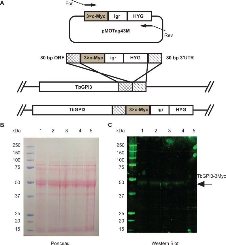
Fig 1. In situ C-terminal tagging of TbGPI3 with 3Myc.
(A) Map of plasmid pMOTag43M [27] used for the in situ tagging of TbGPI3, and a scheme of how the PCR product generated with the indicated forward (For) and reverse (Rev) primers inserts into the 3’-end of the TbGPI3 ORF (checked box) and 3’-UTR (striped box) in the parasite genome to effect in-situ tagging. HYG = hygromycin phosphotransferase selectable marker; igr = α-β tublin intergenic region. (B) Ponceau staining of denaturing SDS-PAGE Western blot shows similar loading and transfer of lysates (corresponding to 5×10⁶ cells) from four in-situ tagged clones (lanes 1–4) and wild type cells (lane 5). (C) The identical blot was probed with anti-Myc antibody. TbGPI-3Myc is indicated by the arrow. The positions of molecular weight markers are indicated on the left of (B) and (C).

