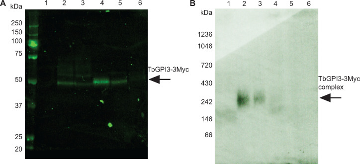
Fig 2. TbGPI3-3Myc is present in complexes in BSF T. brucei.
(A) Aliquots of 2 × 10⁸ cells were harvested and lysed in lysis buffer containing different detergents to assess TbGPI3-3Myc solubilisation. After immunoprecipitation of the supernatants with anti-Myc agarose beads, proteins were eluted with 0.5 mg/mL c-Myc peptide and aliquots were subjected to SDS-PAGE followed by anti-Myc Western blotting. (B) Identical samples were also separated by native-PAGE and subjected to anti-Myc Western blotting. In both cases, lane 1 corresponds to wild type cells lysed with 1% TX-100, as a negative control for anti-Myc blotting, and lanes 2–6 correspond to TbGPI3-3Myc clone1 lysed with 0.5% digitonin, 1% digitonin, 1% TX-100, 1% NOG or 1% DM, respectively.

