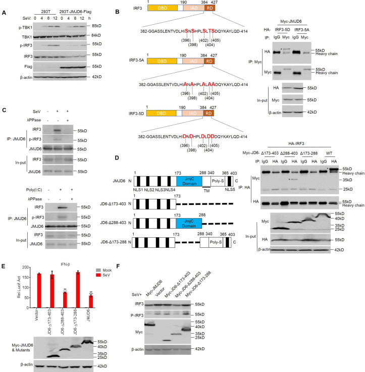
Fig 4. JMJD6 promotes activated IRF3 degradation.
(A) HEK293T cells and HEK293T-JMJD6-Flag cell lines were infected with SeV as indicated, and the phosphorylation levels of IRF3 and TBK1 were analyzed. (B) Diagrams of IRF3 and its mutants. DBD, DNA binding domain; IAD, IRF3 association domain; RD, a C-terminal regulatory domain (left panel). HEK293T cells were transfected with the indicated plasmids, and cell lysates were immunoprecipitated with anti-Myc or IgG antibody followed by immunoblot using anti-HA and anti-Myc antibodies (right panel). (C) Endogenous JMJD6 interacts with IRF3 or p-IRF3 after a viral infection or poly(I:C) stimulation. HEK293T cells were infected with SeV for 12 h. Cells lysates were immunoprecipitated with anti-IRF3 or anti-p-IRF3, and the immunoprecipitates were analyzed by immunoblot with anti-JMJD6 antibody (top panel). Immunoblot of lysates from HEK293T cells stimulated with poly(I:C) for 12 h, analyzed with anti-IRF3 antibody (below panel). (D) Diagrams of JMJD6 and its mutants (Left panel). HEK293T cells transfected with the indicated plasmids and stimulated with poly(I:C) as indicated. The cell lysates were immunoprecipitated with an IgG or anti-HA antibody followed by immunoblots using anti-HA and anti-Myc antibodies. Asterisks represent target proteins (Right panel). (E) HEK293T cells were co-transfected with vector, Myc-JMJD6 or the JMJD6 mutants expressing plasmids and the IFN-β promoter-reporter plasmids for 24 h, and then infected with or without SeV for another 12 h. The luciferase activity was measured with a dual-luciferase assay. Expression of Myc-tagged JMJD6 protein and the mutant proteins was evaluated by Western blotting. (F) HEK293T cells were transfected with the indicated plasmids for 24 h and then infected with SeV for another 12 h. The phosphorylated IRF3 (p-IRF3), total IRF3, Myc-JMJD6 or mutants, and β-actin were detected by Western blotting.

