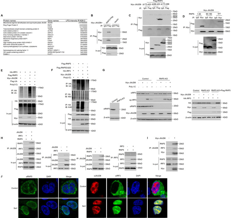
Fig 6. JMJD6 promotes activated IRF3 K48 ubiquitination and degradation via ubiquitin ligase RNF5.
(A) List of JMJD6 interactome based on Label-free quantification intensity. (B) Immunoblot analysis (with anti-Myc or anti-Flag antibodies) of proteins immunoprecipitated (with control IgG or anti-Flag) from lysates of HEK293T cells transfected with the indicated plasmids. The expression of the transfected proteins was analyzed by immunoblot. (C) HEK293T cells were transfected with the indicated plasmids, followed by immunoprecipitation with an IgG or anti-Flag antibody and immunoblotting with anti-Flag or anti-Myc antibodies. (D) HEK293T cells were transfected with the indicated plasmids, followed by immunoprecipitation with an IgG or anti-Myc antibody and immunoblotting with anti-Flag or anti-Myc antibodies. (E) Immunoblot analysis (with anti-Ub) of proteins immunoprecipitated (with anti-HA) from lysates of HEK293T cells transfected for 24 h with various combinations of plasmids (upper panels) and stimulated with poly(I:C) in the presence of MG132. The expression of Flag-RNF5, Myc-JMJD6, or HA-IRF3 was determined by immunoblot with the indicated antibodies (lower panels). (F) Immunoblot analysis (with anti-ubiquitin or antibody to K63-linked or K48-linked polyubiquitin (K63-ub or K48-ub, respectively); top) of proteins immunoprecipitated (with anti-HA) from lysates of HEK293T cells transfected for 24 h with various combinations of plasmids (upper panels) and stimulated with poly(I:C) in the presence of MG132. The expression of Flag-RNF5, Flag-RNF5C42S, Myc-JMJD6, or HA-IRF3 was determined by immunoblot with the indicated antibodies (lower panels). (G) Immunoblot analysis of RNF5 KO efficiency in HEK293T cells (Left panel). Immunoblot analysis of RNF5 KO cells transfected with Myc-JMJD6 or vector and secondarily transfected with poly(I:C), analyzed with anti-IRF3, anti-RNF5, and anti-Myc antibodies (Middle panel). The rescue experiment by ectopically expressing full-length of RNF5 in RNF5-knockout cells (Right panel). (H) In vitro interaction analysis of JMJD6 with RNF5 or IRF3 using in vitro-translated JMJD6, RNF5, and IRF3. JMJD6, RNF5, and IRF3 were obtained by in vitro transcription and translation. Far-left panel, the interaction between JMJD6 and RNF5 or IRF3 was assayed by mixing JMJD6 and RNF5 or IRF3, followed by IP with JMJD6 antibody. Left panel, the interaction between JMJD6 and IRF3, followed by IP with JMJD6 antibody. Right panel, the interaction between JMJD6 and RNF5, followed by IP with JMJD6 antibody. Far-right panel, the interaction between IRF3 and RNF5, followed by IP with IRF3 antibody. (I) Immunoblot analysis of both IRF3 and RNF5 in a JMJD6-immunoprecipitated complex upon virus infection. HEK293T cells were transfected for 24 h with or without Myc-JMJD6 in the presence of SeV infection. (J) Fluorescent images of HEK293T cells stimulated with or without SeV (MOI of 1) for 12 h. DAPI-stained nuclei are shown in blue. Left panel, RNF5 was detected with green fluorescence. Right panel, JMJD6 was detected with red fluorescence, and IRF3 was detected with green fluorescence.

