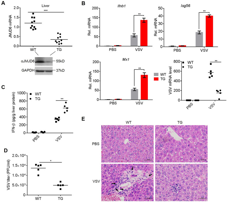
Fig 7. JMJD6 deficient mice are more resistant to VSV infection.
(A) The knockdown efficiency of TG in mouse livers was detected by qPCR (top) and Western blotting (below). (B) qPCR analyses of the levels of the Ifnb1, Isg56, and Mx1 mRNAs and VSV load in the livers of WT and TG mice injected intraperitoneally with PBS or VSV for 48 h. (C) ELISA analysis of IFN-β production in the liver from WT and TG mice intravenously infected with VSV for 48 h. The lowercase letter "g" is the symbol for the gram. (D) The VSV titers of liver from WT and TG mice were analyzed by the standard plaque-forming unit assay. (E) Images of H&E staining of livers sections from the mice. Inflammatory cells are indicated by a black arrowhead.

