Fig. 4.
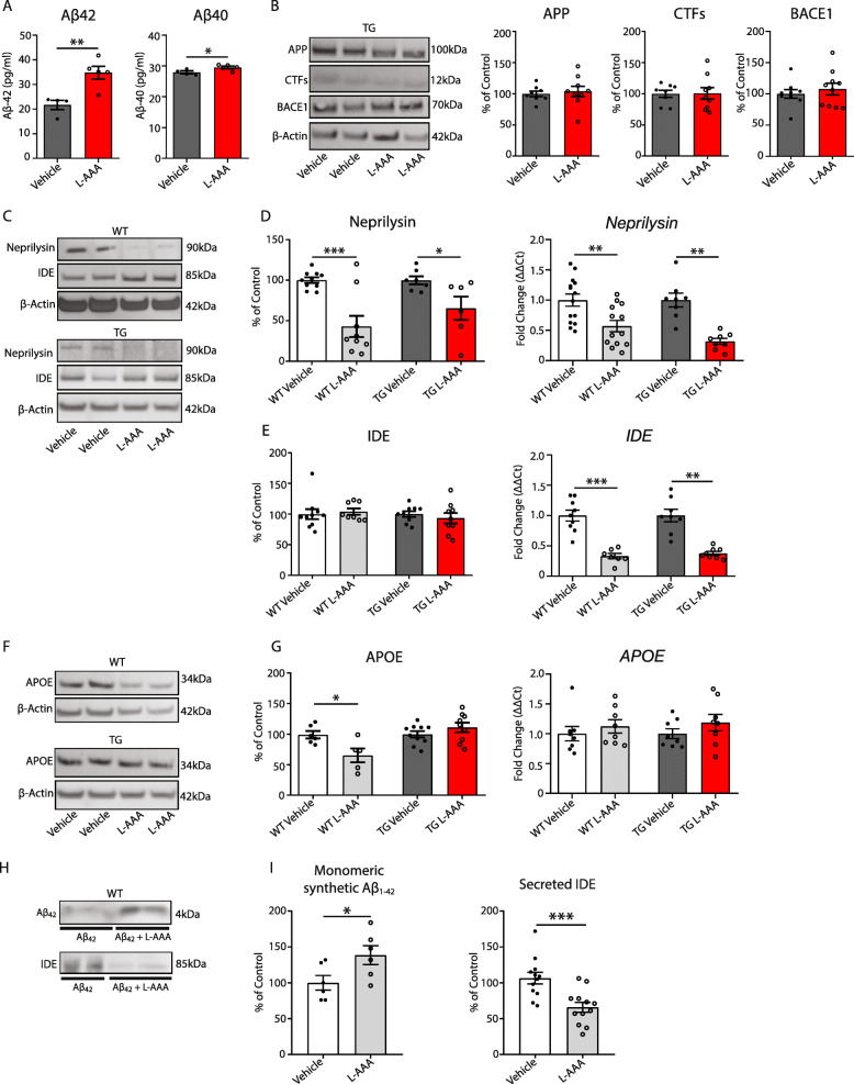
Loss of astrocytes affects Aβ clearance mechanisms. a Quantification of Aβ subtypes by ELISA in 5XFAD OBCSs homogenates (n=4–5). b Quantification and representative Western blot of APP, CTFs, and BACE1 expression in 5XFAD OBCS homogenates (n=6–10). c Representative Western blot of Neprilysin and IDE protein expression in WT and 5XFAD treated with vehicle or L-AAA. d Quantification of Neprilysin protein and gene expression in WT and 5XFAD treated with vehicle or L-AAA (n=6–12). e Quantification of IDE protein and gene expression in WT and 5XFAD OBCSs treated with vehicle or L-AAA (n=6–12). f Representative Western blots and g quantification of ApoE protein and gene expression in WT and 5XFAD OBCSs OBCSs treated with vehicle or L-AAA (n=5–10). h Representative Western blot of synthetic human Aβ1-42 and IDE in conditioned media from WT OBCSs treated for 24h with L-AAA or vehicle i quantification of Aβ42 by Western blot (n=12–18) and quantification of IDE protein expression in conditioned media from WT OBCSs treated for 24 h with synthetic human Aβ1-42 with vehicle or L-AAA (n=12–18). Values shown in graphs represent the mean value ± SEM. Statistical analysis included a Student’s t test, *P<0.05; **P<0.01; ***P<0.001

