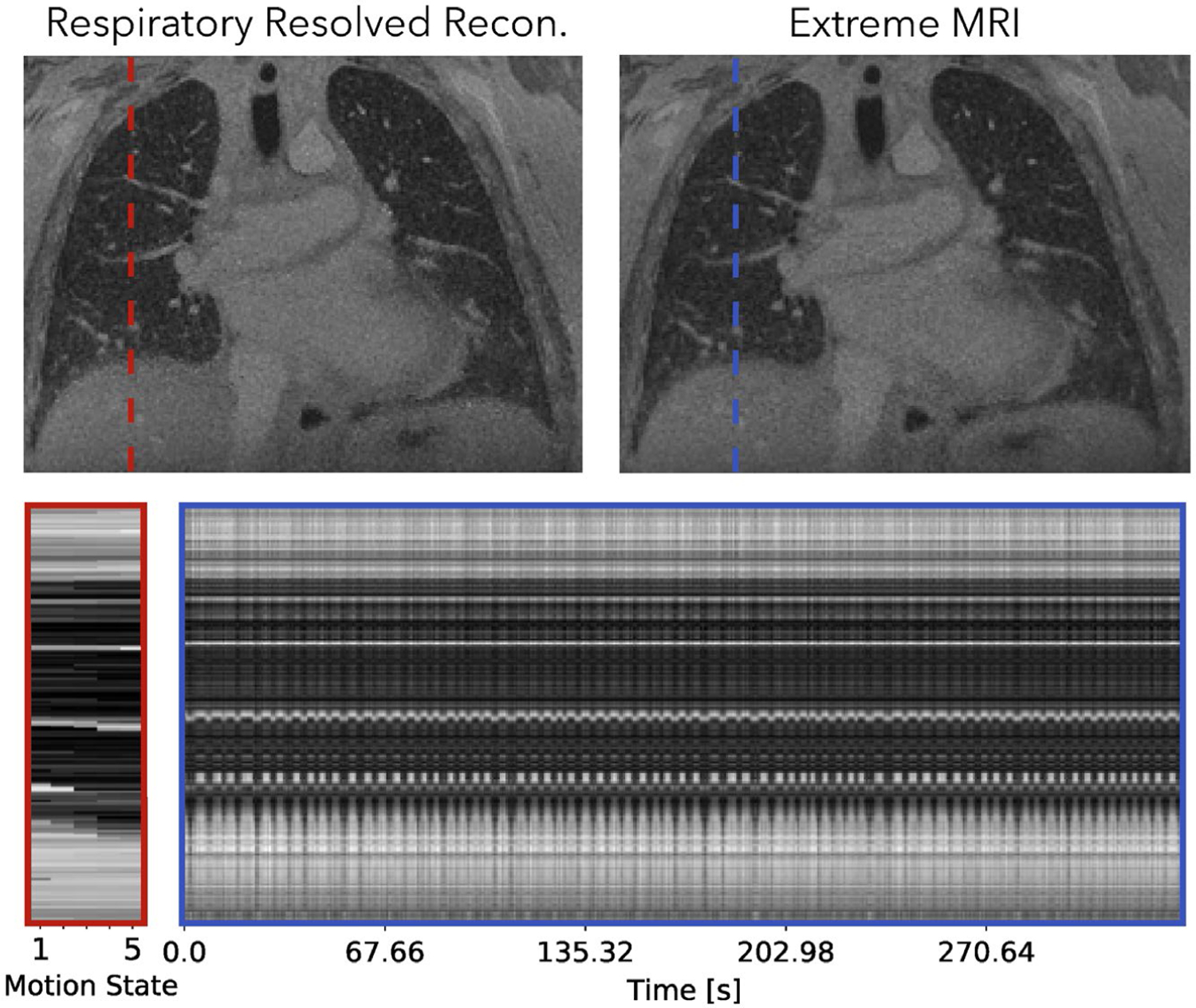
FIGURE 9.

Comparison of the proposed method with the respiratory-resolved reconstruction of the first lung dataset. Dynamics can be seen more clearly in Supporting Information Videos S11 and S12. From the cross section over time and Supporting Information Video S11, regular breathing with slight variable rates can be observed. Overall, Supporting Information Video S11 of the proposed reconstruction shows temporal flickering artifacts. Looking at each frame individually, the proposed reconstruction shows similar image quality and sharpness as the respiratory-resolved reconstruction for the expiration phase. For other phases, the respiratory-resolved reconstruction is slightly sharper near the diaphragms
