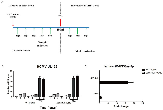
Figure 7.
hcmv-US33as-5p is expressed in both viral latency and reactivation. (A) A schematic diagram depicting in vitro models of latent infection and viral reactivation. THP-1 cells were infected with WT or ΔmiRNA HCMV (MOI = 10) and maintained for 10 days in the culture medium. Then, the THP-1 cells were differentiated into macrophages (d-THP-1) by stimulated with TPA in order to render the cells permissive to HCMV infection and further maintained in the medium for another 5 days. (B) The expression of viral UL122 at 1, 3, 5, 7, and 9 dpi and 1, 3, and 5 days after TPA stimulation was detected by qPCR. (C) The expression of hcmv-US33as-5p at 9 dpi and 3 days after TPA stimulation was determined by qPCR.

