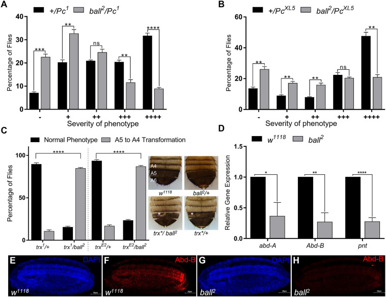
FIGURE 2.
ballchen mutation exhibits trxG like behavior. (A,B) Ballchen mutant flies (ball2) were crossed to two different alleles of Pc (Pc1 and PcXL5). Pc alleles (Pc1 and PcXL5) crossed to w1118 flies were used as control crosses. Heterozygous Pc/+males, Pc1/+ (A) and PcXL5/+ (B), from control crosses exhibit strong extra sex comb phenotype. In contrast, ball2 strongly suppressed the extra sex comb phenotype in both ball2/Pc1 (A) and ball2/PcXL5 (B) male flies. 200 male flies were analyzed for each cross and data shown represents two independent experiments. Male flies were categorized according to the severity of extra sex comb phenotype. These categories are: –, no extra sex combs; +, 1–2 hairs on 2nd leg; ++, more than three hairs on 2nd leg; +++, more than 3 hairs on 2nd leg and 1–2 hairs on 3rd leg; ++++, strong sex combs on both 2nd and 3rd pairs of legs as described previously (Tariq et al., 2009). (C) ball2 mutant flies were crossed to two different alleles of trx (trx1, trxE2). A cross between trx mutants and w1118 served as a control. Males from the resulting progeny were scored for A5 to A4 transformation (loss of pigmentation in A5, marked by asterisk), a known trx mutant phenotype. trx/+heterozygotes from the cross of w1118 with trx mutants were used as control. Compared to the control, ball2/trx1 and ball2/trxE2 showed a higher percentage of flies with A5 to A4 transformation, indicating a strong enhancement of trx mutant phenotype. Representative images of w1118, ball, and trx heterozygous mutants and ball/trx double mutants are shown. The expressivity of A5 to A4 transformation phenotype of trx*/ball2 was comparable to trx*/+. All crosses were carried out in triplicates and independent t-tests were performed for analyzing each category (*p ≤ 0.05, **p ≤ 0.01, ***p ≤ 0.001, or ****p ≤ 0.0001). (D) Significantly low levels of abd-A, Abd-B and pnt expression was detected through qRT-PCR in homozygous ball2 embryos when compared with w1118 embryos. Independent t-tests were performed for each gene analysis (*p ≤ 0.05, **p ≤ 0.01, ***p ≤ 0.001, or ****p ≤ 0.0001). (E–H) Immunostaining of stage 15 embryos with Abd-B antibody is shown in w1118 (E,F) as well as homozygous ball2 (G,H) embryos. As compared to w1118 (F), ball2 embryos showed strongly diminished Abd-B (H) expression.

