FIGURE 3.
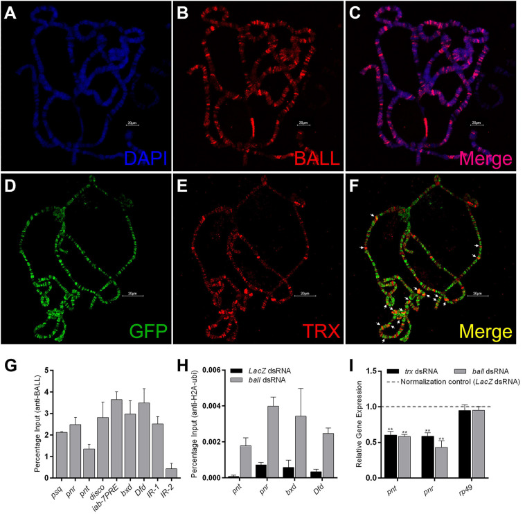
BALL co-localizes with TRX at the chromatin and antagonizes PcG. (A–C) Polytene chromosomes from third instar larvae stained with DAPI (A) and BALL antibody (B). (D–F) Third instar larvae expressing GFP-tagged BALL were stained with GFP (D) and TRX (E) antibodies. Co-localization of BALL and TRX is clearly seen at several loci [(F), white arrows]. (G) Strong enrichment of BALL at non-homeotic (psq, pnr, pnt, and disco) and homeotic (iab-7PRE, bxd, Dfd) trxG targets was observed by ChIP from wild-type Drosophila S2 cells using BALL antibody. IR-1 and IR-2 are intergenic regions not bound by TRX and served as controls (Papp and Müller, 2006). (H) ChIP from BALL depleted cells showed enhanced H2AK118ub1 levels at non-homeotic (pnt, pnr) and homeotic (bxd, Dfd) targets of trxG as compared to cells treated with dsRNA against LacZ. (I) Using qRT-PCR, relative gene expression of pnt and pnr was analyzed in cells after ball or trx knockdown. LacZ dsRNA treated cells were used as normalization control (dashed line) and the expression of pnt and pnr in ball and trx depleted cells was plotted. BALL depleted cells showed a significantly decreased expression of pnt and pnr similar to the cells with trx knockdown. The mRNA levels of a ribosomal protein, rp49, served as a negative control. Independent t-tests were performed for analyzing relative expression of each gene (*p ≤ 0.05, **p ≤ 0.01, ***p ≤ 0.001, or ****p ≤ 0.0001).

