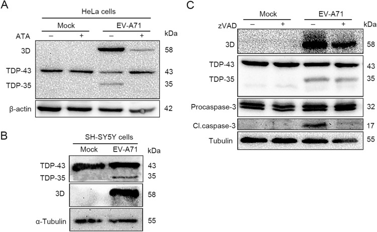
Fig. 1.
TDP-43 is cleaved in the cells infected with EV-A71. A–B HeLa cells and SH-SY5Y were cultured in 12-well plates to 60%–70% confluence and infected with EV-A71 at 1 × 10⁴ TCID50 for 24 h. TDP-43 proteins were analyzed by Western blotting. HeLa cells were cultured in 12-well plates and infected or mock-infected EV-A71 for 24 h in the medium containing 10 µmol/L ATA (aurintricarboxylic acid, 3D inhibitor), the inhibitor of 3D polymerase of enteroviruses (A) or 20 μmol/L pan-caspase inhibitor Z-VAD-FMK (C). Cell lysates were subjected to Western blot analysis.

