Fig. 5.
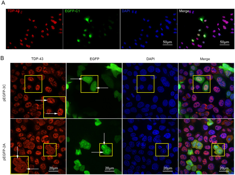
The cytoplasmic translocation of TDP-43 is caused by protease 2A of EV-A71. HeLa cells were transiently transfected with pEGFP-C1 (A), pEGFP-2A (B), and pEGFP-3C (B), respectively, for 24 h. Cells were harvested for immunofluorescence staining using anti-TDP-43 antibody (Alexa-fluor-594). Cell nuclei were stained with DAPI. Fluorescence images were taken by fluorescence microscope (A) and CV1000 confocal system (B). Yellow squares in the upper panel of B indicate the cells expressing EGFP-3C. Yellow squares in the lower panel of B indicate the cells expressing EGFP-2A. The larger square in the upper right or lower right panel of B is the magnified image enclosed by the smaller square in the same panel. The distribution of TDP-43 was indicated by arrows in the upper right and lower right panels of B. Arrows in the upper-middle of B indicate the nuclei of the cells expressing EGFP-3C, while arrows in the lower-middle of B indicate the cytoplasm of the cells expressing EGFP-2A.

