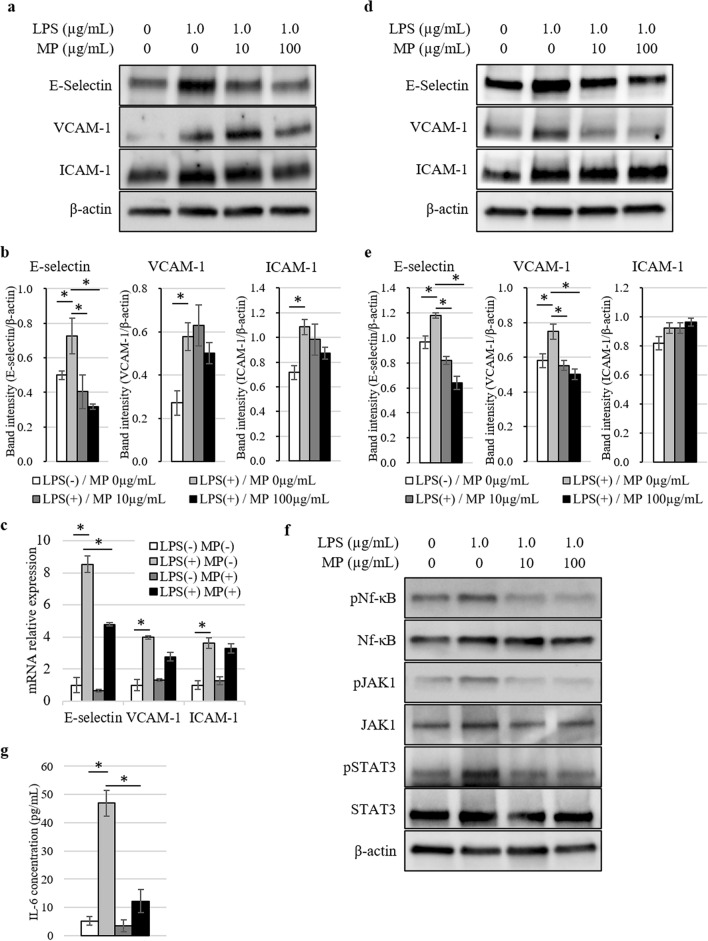
Figure 1.
Analysis of adhesion molecules expressed in vascular endothelial cells stimulated by LPS and MP. (a) Protein expression levels of E-selectin, VCAM-1, and ICAM-1 in HUVEC at 6 h after stimulation analyzed by western blotting. Blot was cut horizontally and immunoblotting was performed for each section. (b) Quantification of E-selectin, VCAM-1, and ICAM-1 band intensity normalized to β-actin in HUVEC. Data are means ± standard error (n = 3, each group); *P < 0.05. (c) Induction of SELE, VCAM1, and ICAM1 gene expression in HUVEC 6 h after stimulated by 1.0 µg/mL of LPS and 100 µg/mL of MP was analyzed by quantitative real-time PCR. Data are normalized relative to ACTB mRNA levels. Data are means ± standard error (n = 3, each group); *P < 0.05. (d) Protein expression levels of E-selectin, VCAM-1, and ICAM-1 in HHSEC at 6 h after stimulation analyzed by western blotting. Blot was cut horizontally and immunoblotting was performed for each section. (e) Quantification of E-selectin, VCAM-1, and ICAM-1 band intensity normalized to β-actin in HHSEC. Data are means ± standard error (n = 3, each group); *P < 0.05. (f) The phosphorylation of Nf-κB, JAK1, and STAT3 was analyzed by western blotting with antibodies targeting pNf-κB/Nf-κB, pJAK1/JAK1, and pSTAT3/STAT3 at 30 min after the stimulation. Blot was cut horizontally and immunoblotting was performed for each section. (g) ELISA analysis of IL-6 concentration levels in conditioned medium 6 h after stimulated by 1.0 µg/mL of LPS and 100 µg/mL of MP. Data are means ± standard error (n = 3, each group); *P < 0.05.

