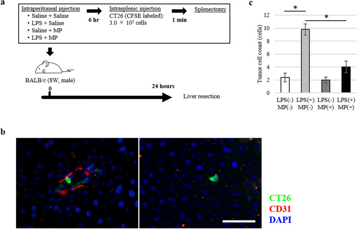
Figure 4.
Suppressive effects of MP on tumour cell hepatic binding in LPS-induced murine hepatic metastasis models. (a) Mouse colon cancer cell line (CT26) hepatic metastasis model; male BALB/c mice (8 weeks old) were treated with saline/saline (control), LPS/saline, saline/MP, or LPS/MP. Six hours later, 3.0 × 10⁵ CT26 cells were injected into the inferior pole of the spleen, followed by splenectomy 1 min later. After 24 h, the liver was resected for analysis. (b) Representative images of CT26 tumour cells adhered to the endothelial cells (left) and transmigrated into the liver (right). CT26 tumour cells are labeled by CFSE fluorescence (green). Each protein was co-stained with anti-CD31 antibody to define vascular endothelial cells (red). Nuclei are stained with DAPI (blue). Scale bars are equal to 50 µm. (c) Number of CT26 tumour cells binding to the liver following pretreatment with LPS and MP. Data are means ± standard error (n = 5, each group); *P < 0.01.

