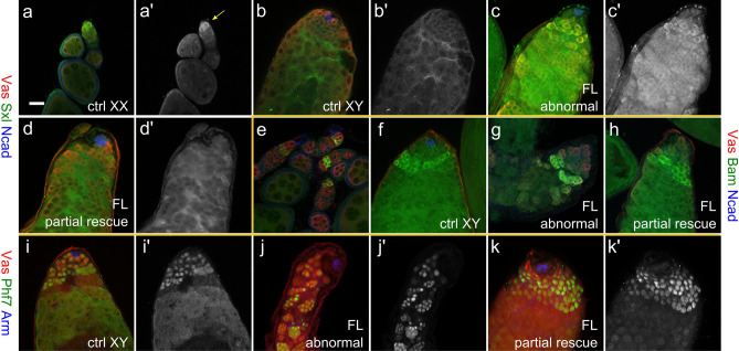
Figure 3.
Expression of sex-specific markers in the pseudotestes. (a–d), Sxl staining in wild-type ovaries (a), wild-type testis (b), and abnormal (c) and partial rescue (d) type pseudotestes of UAS-Phf7.FL, Δtra/tra1, nos-Gal4. Vasa is in red, Sxl in green, and N-cadherin in blue. (a′–d′) display just the Sxl signals alone. The yellow arrow in A′ indicate the germline stem cells in an ovariole that stain Sxl clearly. (e–h), Staining of Bam in wild-type ovary (e), wild-type testis (f), and abnormal (g) and partial rescue (h) type pseudotestes of UAS-Phf7.FL, Δtra/tra1, nos-Gal4. Vasa is in red, Bam in green, and N-cadherin in blue. (i–k), Phf7 expression in wild-type testes (i), and abnormal (j) and partial rescue (k) type pseudotestes of UAS-Phf7.FL, Δtra/tra1, nos-Gal4. Vasa is in red, Phf7 in green, and Armadillo in blue. (i′–k′) display the Phf7 channel alone.

