Figure 5.
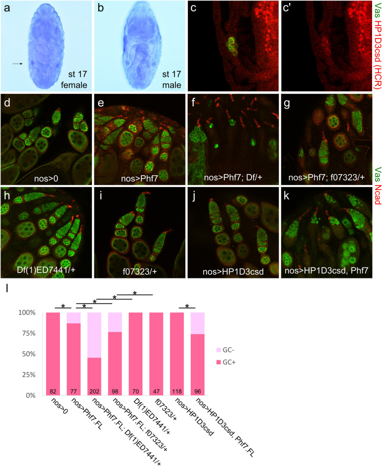
Genetic interaction between Phf7 and HP1D3csd on the female germline loss caused by Phf7 overexpression. (a,b), in situ hybridization of stage 17 embryos with an HP1D3csd probe. (a), Female embryo; arrow indicates position of gonad on one side. (b), Male embryo. (c), HCR staining of HP1D3csd transcripts in an unsexed stage 17 embryo. Colocalization with the Vas (green) signal indicate the HP1D3csd signals are in germ cells. (c'), HCR signal alone. (d–k), Ovaries of various genotypes stained with Vasa (green) and N-cadherin (red). (d), nos-Gal4/ + , (e), nos-Gal4/UAS-Phf7.FL, (f), Df(1)ED7441/ + ; nos-Gal4/UAS-Phf7.FL, (g), HP1D3csdf07323/ + ; nos-Gal4/UAS-Phf7.FL, (h), Df(1)ED7441/ + , (i), HP1D3csdf07323/ + , (j), nos-Gal4/UAS-HP1D3csd.HA, (k), nos-Gal4/UAS-HP1D3csd.HA, UAS-Phf7.FL. (l), Quantitation of the fraction of ovarioles that contain no germline in different genotypes. Sample sizes and genotypes are indicated on the bottom of the bars. Dark and light pink portions of the bars denote the fraction of ovaries with and without germline. * indicates P < 0.05 with chi-square tests.

